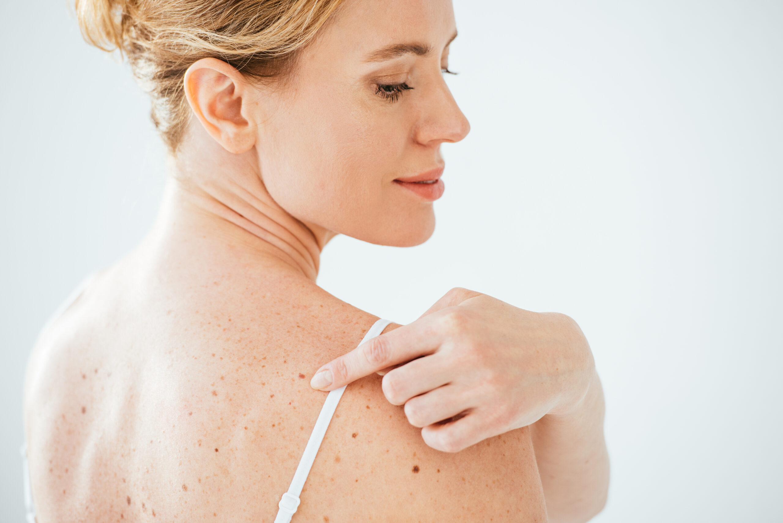
Mole Mapping
Mole mapping – also called Total Body Photography (TBP) – is a process that uses photography to capture detailed images of the whole body and skin lesions over time so these can be compared side-by-side and changes such as size, colour, and structure can be determined early.
Total Body Photography is safe, non-invasive and painless. It aims to detect melanoma early. That’s when treatment can be 95 percent effective, leading to a high survival rate. By identifying suspicious or evolving changes, another benefit of mole mapping is that it can provide a guide to which lesions need removing, thereby reducing the number of biopsies and subsequent scars.
Why is mole mapping important?
Melanoma is a dangerous skin cancer with limited effective treatment once it spreads. It often starts with changes in a previously existing mole or appears as a new mole, which sometimes can be overlooked on the background of a very “moley” skin. The good news is regular assessment and comparison gives us the opportunity to track new changes and make early treatment interventions.
Who should have Total Body Photography?
You should consider doing periodic total body photography if:
-
- 1. You have had a personal or family history;
-
- 2. You sunburn very easily;
-
- 3. You have numerous moles (usually >30) especially in areas difficult to monitor by yourself, such as your back, neck, and back of thighs;
-
- 4. You have had an atypical mole or other cancers removed from the skin.
How is it done?
This is a head-to-toe inspection and photography. It is best to wear loose clothing to your appointment and you may be offered to change into a hospital gown for your modesty. Photographs of the body will be taken in sections and close-up photos and inspection of suspicious moles will also be taken. Areas constantly exposed to the sun are likely to get more damaged, though photos will be taken of the whole body. You may choose not to have your buttocks or genitals examined.
What happens after the photography?
You will be provided copies of your photos for your reference to keep.
If a lesion is suspected to be cancerous, you will be advised to see your family physician to arrange a biopsy. Lesions that are concerning for atypical appearance are reviewed after six to 12 months for signs of changes.
- Treatment time : 20-40 minutes
- Anesthesia : None
- Recovery Time : None
- Treatment and Result : Cancerous-suspect or evolving lesions are biopsied. Other lesions are reassessed after 6-12 months.
